The fundus exam is a painless, fast and simple test that is very useful to detect eye problems. Ideally, a person should have it done once a year, especially after the age of 50. Ideally, the fundus exam should be part of anyone’s regular medical check-ups, but this is not always the case. It is a simple test that allows you to detect common eye problems, even before they generate symptoms.
IS THE FUNDUS SCAN PAINFUL?
It is not a painful test, during the fundus test, the ophthalmologist will ask you to fix your eyes on a point and not blink for a few seconds. This test is routine and painless, so you don’t need to worry. It lasts no more than ten minutes and you can go home after the scan.
However, if your pupil has dilated, as the eye will still be dilated for a while and the vision will be somewhat blurred, it is advisable to always be accompanied to the consultation and use sunglasses when going outside. Our eyes will be very sensitive to light and we may be dazzled. It is also prudent to wait for us to see correctly before driving or using machinery for whose use greater visual acuity is needed.
The fundus exam is a technique that makes it possible to look inside the eyeball. This makes it possible to detect the presence of any abnormality in vision. It also serves to follow the evolution of some pathologies such as hypertension and diabetes. This is considered the most routine test in ophthalmology. It is painless and does not cause discomfort beyond pupil dilation, which causes blurred vision for a few hours. The fundus examination allows to observe the state of the optic nerve, blood vessels and retina.
What is a fundus exam?
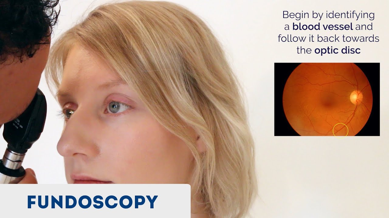
The fundus exam is an examination test performed by the ophthalmologist to look at the back and inside of the eye. It is also known as an ophthalmoscopy or funduscopy. This technique allows you to see the deepest part of the eye through the pupil. With this examination it is possible to visualize areas such as the macula, the retina, the optic disc and the transparent media that are: the cornea, the lens, the vitreous humor and the aqueous humor. This technique can also track eye problems such as glaucoma, macular degeneration, eye cancer, optic nerve problems, and eye injuries.
Types of Fundus Examination
The fundus exam is done with a device called an ophthalmoscope. It is composed of a series of crystals and mirrors that illuminate the retina, without light being reflected. This allows you to observe everything clearly, without any flashes.
Depending on the type of ophthalmoscope used, there are three types of fundus examination:
- Direct ophthalmoscopy. It is the simplest technique. It allows to obtain the image of a single eye, which is one-dimensional; that is, flat.
- Indirect ophthalmoscopy. It is a more complex technique that makes it possible to obtain the image of the two eyes.
- Indirect ophthalmoscopy with slit lamp. It is the most complex of all and allows to obtain a three-dimensional image of both eyes.
At present, other more advanced techniques are also applied, such as digital photographs of the fundus of the eye that allow detecting the evolution of the retina in certain pathologies. Also fluorescein angiography, which makes it possible to see in detail the blood vessels.
What is a fundus exam for?
The main utility of the fundus examination is that it allows to identify serious eye diseases in their initial phases. The test also makes it possible to track anomalies that have been identified. With this test you can detect eye diseases, especially those concerning the retina, other discomfort in the eyes such as those that cause headache due to vision problems, and other head injuries.
This test allows to diagnose, among others, the following disorders:
- Melanoma of the eye.
- Diabetic retinopathy.
- Glaucoma.
- Age-related macular degeneration.
- Retinal detachment. It is a separation of the retina, more common in myopic people.
- Retinal thrombosis. Obstruction of any of the veins that carry blood to the retina.
- Macular degeneration. It causes central and acute vision to be lost.
- Glaucoma. It is a loss of peripheral vision due to damage to the optic nerve. If not treated in time, it can cause blindness.
- Ocular melanoma. It is a type of cancer.
- Diabetic retinopathy. It is damage to the blood vessels of the retina, caused by diabetes.
The most interesting thing is that with the fundus examination non-ocular diseases are also detected, such as hypertension and diabetes. It is believed that in the future it will make it possible to diagnose, even, neurodegenerative diseases.
TYPES OF FUNDUS EXAM
Fundus directly
The ophthalmologist uses a device the size of a flashlight that emits a powerful light and that inside has several lenses that increase the size of the area to be studied.
Fundus indirectly
For its part, the indirect consists of a device that includes a small microscope with light, which is known as a slit lamp, which allows to visualize the fundus of the eye with a greater amplitude of size.
When a fundus is made indirectly, the patient has to be seated, in a comfortable position, and place the head on a device where he holds the chin and forehead, in this way, he does not move to be able to make the fundus reliably. Depending on your case and your symptoms, the ophthalmologist will use the most appropriate one.
How is a fundus exam performed?
- To perform a fundus it is necessary to dilate the pupil.
- When you arrive at the fundus examination consultation, the first thing you do is apply a few drops to your eye. These can sting a little and their function is to dilate the pupil little by little. The process can take anywhere from a few minutes to an hour.
- When dilation is deemed to be at the sweet spot, the patient is asked to come to the office and the lights are turned off. If the direct or indirect technique is used, the person will have to sit in a reclining chair or lie on a stretcher.
- The doctor will ask the person to fix their eyes on a specific point and keep it that way for a moment. In the direct technique, you will look at each eye separately. In the direct, you will examine both eyes at once; in the latter case, the ophthalmologist will use a device similar to a miner’s helmet.
- If the fundus examination is done with a slit lamp, the patient will have to sit in a chair and rest the chin on a metal device. You will also need to fix your gaze on a point to make the exam possible. In all cases, the exam lasts only a few minutes.
Contraindications to this test
- The fundus exam rarely leads to complications. Only in very few cases can there be allergy to the drops that are applied to the eyes. This causes more itching than normal and redness of the eyes.
- There are no absolute contraindications, but ophthalmologists take special caution in cases of people with glaucoma, as the drops can cause an increase in intraocular pressure and lead to an acute attack.
- Special care should also be taken when a person has cataracts, as the lens may be thickened. Either way, the doctor will assess the situation and take the necessary measures so as not to generate risks to the patient.
- An examination that requires accompaniment by the patient
- After the fundus examination is performed, there may be some discomfort. The pupil will have dilated and will not return to normal until after a few hours. Therefore, there will be blurred vision and you will feel discomfort in the sunlight.
- During the following hours, it will not be possible to perform actions such as reading or any activity that requires sharp vision. For the same reason, it is advisable that the person is accompanied to this exam, so that he does not have difficulties to return home.